Table of Contents
Artificial intelligence and machine learning are on the rise, and healthcare is among the industries where AI/ML can drive changes to drastically reinvent approaches to diagnostics, patient care, hospital management, etc.
Artificial intelligence is always tied to data, and for data-intensive sources like medical images, it can power up a new level of analytical depth and help radiologists process images more efficiently and precisely than ever.
The workload of radiologists increases exponentially: in some cases, average radiologists need to read a medical image within 3-4 seconds to complete the amount of work they have in an 8-hour day.
Of course, such an intensity won’t reflect positively on healthcare professionals’ mental state and concentration, which eventually leads to low diagnostic accuracy, additional stress for patients, and fast burnout for healthcare specialists.
Considering these conditions, medical imaging machine learning is a key to relieving radiologists from excessive workload, but the true potential of these technologies goes far beyond and allows for detecting abnormalities in medical images that the human eye may miss or misinterpret.
Let’s contemplate some practical cases of artificial intelligence and image recognition software being applied to the field of medical image analysis.
Brain tumors

The treatment for a brain tumour depends on its type. To this day, the main course of action for a patient with such a diagnosis includes surgery, during which the surgeon removes as much of the tumour mass as possible.
Then, the sample is analysed to determine whether it is a benign or malignant tumour and where the margin is between healthy tissues and tumour cells. The conventional analysis takes a certain amount of time, and the surgeon and patients are bound to wait for this period to understand their next steps.
Add AI and SRH (stimulated Raman histology) to the equation, and you will be able to obtain accurate results in three minutes within the operating room. While surgery is the most efficient way to find answers, it is still pretty invasive.
There is also always a risk of a surgeon removing too much healthy tissue, which might lead to further neurological problems. Hence, researchers decided to go further and develop a deep-learning model that will be able to detect and classify brain tumours using solely MRIS.
In their study, researchers increased the accuracy of diagnosis on an MRI basis by 12 percentage points, up to 75.5%, which shows that deep learning methods have the potential to be an effective tool for brain tumour detection and classification.
Neurological diseases
MRI is the major tool used for the diagnosis of neurological conditions like Alzheimer’s disease, multiple sclerosis, amyotrophic lateral sclerosis, etc. An MRI scan allows doctors to assess the condition of the patient by monitoring brain lesions, their growth, or shrinkage.
However, slight changes might not be noticeable to the eye, even to the most experienced professional, leaving patients and their families in oblivion for a pretty long period of time. And here is where artificial intelligence can strengthen radiologists by helping to detect and quantify changes in the patient’s brain and allowing early detection of developing disease and faster diagnosis.
Within the last few years, researchers have presented algorithms for detecting Alzheimer’s and multiple sclerosis.
Breast cancer

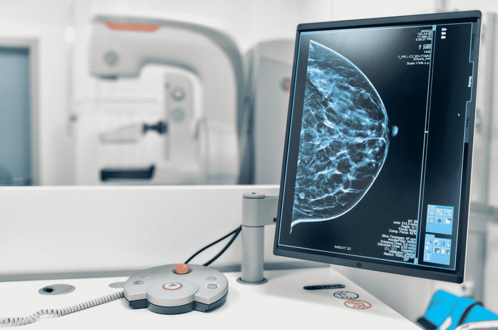
Scientists work on developing a machine learning algorithm that will show sufficient accuracy to be implemented into clinical practices. Unfortunately, the latest studies on the usage of AI for mammography screening programs show that health care professionals are still more efficient in detecting breast cancer than AI, however, the potential is there, and it has every chance to become a powerful tool for efficient diagnostic in the future.
At the same time, for ultrasound exams, a less common but more available type of screening, AI has already shown better performance: radiologists were able to increase the accuracy of their diagnosis up to 96% when using AI. However, there is still a need for further trials, as researchers used only historical data from past exams in their analysis ,and real-life conditions might make their adjustments.
Cardiovascular issues
Cardiovascular diseases are the main cause of death worldwide, which only pushes scientists to pursue the implementation of artificial intelligence for faster and more accurate diagnosis. Hence, more and more researchers work on AI algorithms dedicated to these purposes, using different approaches to diagnostics.
One of the already developed and implemented tools is the algorithm, which can analyse CRM scans for any non-congenital cardiovascular disease. After being trained on almost 2,000 scans and fed a dataset with an additional 1,200 scans, the solution needs 20 seconds to run the analysis, which is a significant improvement compared to the 13 minutes a human reader spends on a single image.
At the same time, the project’s authors claim that such speed doesn’t jeopardise the precision of the scanning. Another worthy example is an AI solution designed to create a 3D model of arteries from CT scans in order to avoid invasive exams while diagnosing coronary artery disease (CAD).
Among cardiovascular diseases, CAD is a leading cause of disease, and historically, the only way to detect it was cardiac catheterisation. This invasive procedure poses the risk of complications and creates additional stress for the patient.
In turn, an AI-powered 3d modelling tool allows for making the diagnostic process more patient-friendly. Apart from diagnostics, researchers are trying to leverage AI for risk prediction. Currently, such solutions are still underdeveloped for clinical practice, but with ongoing research, there are great chances of seeing AI solutions implemented to predict and prevent cardiovascular diseases.
For example, in this early study, researchers trained a deep learning algorithm to predict a 10-year risk of heart disease by analysing a single X-ray. And while the tool still lacks precision, it shows promise to become an efficient alternative to traditional risk estimation and identify high-risk patients who could otherwise go unnoticed.
Musculoskeletal injuries

AI can significantly contribute to the detection of musculoskeletal issues, particularly bone fractures. There are several reasons why fractures might be unnoticed by clinicians (overload that increases the chance of error, prioritisation of other clinical concerns like internal bleeding or organ injury, lack of radiologists in a facility, etc.).
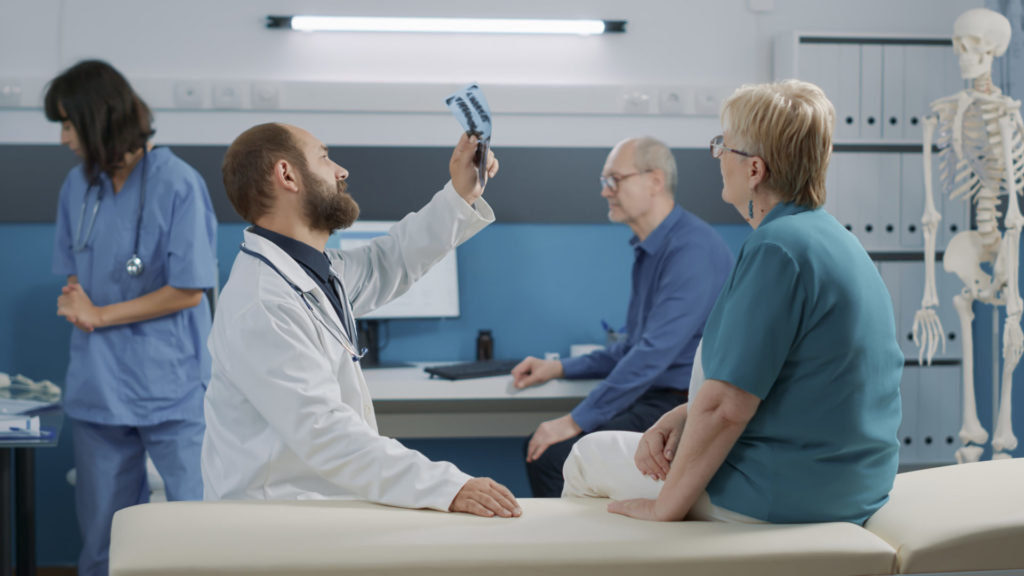
Whatever the case is, artificial intelligence can aid healthcare professionals and improve the fracture diagnosis success rate or act as a “second reader” to ensure accuracy. However, like many other AI-powered medical imaging tools, it is still in the early stages of development and requires improvements before being implemented in the clinical environment for fracture detection.
At the same time, AI can be used to analyse medical imaging data to detect other musculoskeletal injuries and issues, like identifying the problems with joint replacement devices, more efficient assessment of rotator cuff tears, etc.
Second opinion
While the majority of artificial intelligence algorithms might not yet be sufficiently trained to be the only basis for decision-making, AI for medical imaging can be successfully used as a “second reader” to provide a second opinion for a certain case or to run a cross-validation of clinicians’ diagnoses if there are any doubts.
The relevant algorithms can work for any type of medical imaging (MRI, CT, ultrasound, X-ray images) and become a powerful aid to radiologists who suffer from excessive workloads.
In 2020, in the middle of the COVID-19 pandemic, US researchers created an AI algorithm that allows the detection of COVID by analysing CT images of patient lungs. The value of this study is especially high: at the moment, CT has not been the most common method of detecting COVID, with genetic exams prevailing. While labs have been overwhelmed, CT could become another option for diagnosis, which would increase the percentage of early diagnosis and timely isolation of infected patients.
In this case, AI will strengthen the diagnostic capacities of clinicians by being able to detect the disease during the early course of the illness or by discovering non-specific findings.
Radiation dosage reduction
The process of creating medical images like CT or MRI requires that patients be exposed to radiation. This pushed researchers to study the topic of dosage optimisation to improve patient care and reduce the impact of harmful exposure, which is especially important in paediatrics.
A lot of studies are dedicated to the capacity of deep learning to increase the resolution of CT/MRI scans taken with a lower dosage of radiation than average. Already, some tools can improve the quality of images taken with 10% of the original dose, but the question is whether such images could be trusted and how much information on the image was generated by AI without any comprehensive reasoning behind it.
AI in personalised treatment plans
AI is central in creating personalised treatment plans by analysing individual patient data. Genetic information, medical history, and diagnostic imaging provide valuable information to design treatments tailored to the unique characteristics of each patient. By doing so, AI helps improve the chances of successful outcomes and reduces the need for a trial-and-error approach in treatment.
Enhancing precision medicine accuracy with AI
One of AI’s most impactful contributions to precision medicine is its ability to improve diagnostic accuracy. AI provides a comprehensive view of a patient’s health, allowing for more accurate diagnoses. It also leverages multimodal image classification to cross-reference data from different modalities, ensuring more reliable results and reducing diagnostic errors.
AI in clinical decision support systems
AI-powered clinical decision support systems (CDSS) assist healthcare providers by offering real-time, data-driven insights. These systems analyse vast amounts of patient data and medical literature, presenting clinicians with tailored recommendations. As a result, it supports decision-making in complex cases, improves consistency, and reduces the likelihood of human error.
AI-enabled early intervention in healthcare
AI’s ability to detect early signs of disease is helping healthcare professionals intervene sooner, often before symptoms appear. AI facilitates early intervention, potentially improving long-term outcomes for conditions such as cancer, Alzheimer’s, and heart disease by identifying subtle changes in imaging and searching patient records for risk factors.
Streamlining ALS diagnosis with AI algorithms
AI algorithms are particularly effective in streamlining the diagnosis of conditions like ALS (amyotrophic lateral sclerosis). AI can identify abnormalities that may be difficult for the human eye to detect. Early flagging of suspicious results enables faster, more accurate diagnoses and allows healthcare providers to initiate treatment sooner.
Augmented visualisation in surgery with AI
AI is also transforming the surgical field by providing augmented visualisation, which enhances surgical precision. Through advanced imaging and real-time analysis, AI allows surgeons to view more detailed anatomical structures and plan their procedures with greater accuracy. This not only improves outcomes but also minimises the risk of complications during surgery.
The main challenges behind artificial intelligence and machine learning in medical imaging
AI in medical imaging is a hot topic, however, there are still significant challenges that scientists, AI developers, and medical professionals should address to achieve success in the field.
Generalisability of models
Deep learning models, widely used for medical image analysis, are trained on certain data pieces to predict outcomes when fed raw data. But what if the data you are going to feed to the model differs from the original pieces, e.g., if you decide to use data for patients from another geographical region? Would you be able to trust the algorithm? Will it be able to make the right conclusions?
There is always a chance that an algorithm will show lower accuracy on a dataset different from the one that was used to study the algorithm, and it might be the biggest challenge that stops AI-based medical imaging tools from clinical implementation.
Data acquisition
To create a precise solution, developers need a lot of representative data to train the algorithm. But it is pretty hard to acquire enough data, as medical institutions don’t want to share data easily. And of course, there is also an important question of regulations and privacy that complicates data acquisition.
Lack of infrastructure in hospitals
There is a lack of clinical trials, and the main reason is that the majority of hospitals don’t have the computational infrastructure to track and evaluate the performance of AI algorithms, which makes it pretty hard to run clinical trials and check the deep learning performance in a clinical environment.
In conclusion
The implementation of artificial intelligence for medical imaging has a lot of potential; no wonder it is arousing the interest of researchers. And while some of the solutions are already used in certain medical institutions, AI-based tools are still in the early stages of their development, and their real mass use lies ahead of us.
Still, hospitals and other medical institutions can benefit from AI capacities right now and improve the lives of clinicians and patients. Image recognition can lower the probability of human errors in medical records and help you process some cases of medical images.
AI-based solutions for your medical institution
Enhance the performance of your team with the help of artificial intelligence
Contact usThe most common type of machine learning used for medical imaging purposes is deep learning.
Machine learning is used to recognize patterns: the algorithm computes the image features, that identify the best combination of these features to classify the image or to create some metrics related to a certain image region.
AI-powered medical imaging significantly improves early detection by quickly analysing images for subtle signs of disease that the human eye might miss. Early identification of conditions like cancer or cardiovascular issues can lead to more effective treatment options and better patient outcomes. The key benefit here is its ability to provide fast, reliable insights, enabling doctors to act sooner and potentially save lives.
AI reduces diagnostic errors by leveraging vast amounts of medical data to recognise patterns and anomalies. Once trained on large datasets, AI systems can analyse images with high accuracy, reducing human error caused by fatigue, distractions, or varying skill levels. The result is more consistent interpretations, ensuring that diagnoses are based on the best available evidence rather than subjective judgment.
Multimodal image classification combines data from different imaging techniques—such as X-rays, MRIs, and CT scans—allowing for a more comprehensive analysis. This approach leads to better diagnostic accuracy by providing a fuller picture of the patient’s condition. AI can cross-reference different modalities to detect conditions that may be overlooked when analysing a single image type, enhancing overall diagnostic reliability.
AI streamlines clinical workflows by automating routine tasks, such as image preprocessing, segmentation, and analysis. It can prioritise cases based on urgency or complexity, ensuring that doctors focus on the most critical patients first. AI also integrates with existing healthcare systems, reducing administrative burden and helping medical professionals deliver care more efficiently and effectively.